Homepage
/ Pelvic Female Abdomen Ultrasound : Ultrasound Scanning Of The Pelvis And Abdomen For Staging Of Gynecological Tumors A Review Fischerova 2011 Ultrasound In Obstetrics Amp Gynecology Wiley Online Library
Pelvic Female Abdomen Ultrasound : Ultrasound Scanning Of The Pelvis And Abdomen For Staging Of Gynecological Tumors A Review Fischerova 2011 Ultrasound In Obstetrics Amp Gynecology Wiley Online Library
Pelvic Female Abdomen Ultrasound : Ultrasound Scanning Of The Pelvis And Abdomen For Staging Of Gynecological Tumors A Review Fischerova 2011 Ultrasound In Obstetrics Amp Gynecology Wiley Online Library. What is a female pelvic ultrasound? Pelvic ultrasound is also used during a biopsy to help guide the needle. Ultrasound of the female pelvis. Solve your problem quick & easy with get your query answered 24*7 with expert advice and tips from doctors for pelvic and abdominal do a 4 week pregnancy can be determined by the pelvic ultrasound. A pelvic ultrasound is a test that uses sound waves to make pictures of the organs inside your pelvis.
Is pelvic and abdominal ultrasound your major concern? If your doctor orders a pelvic ultrasound exam, images can be captured in two different ways: Ultrasound of the female pelvis. The test can be done in two ways Structures pictured on pelvic ultrasound:
Pelvic Ultrasound Scans For Women In London Ultrasound Services from ultrasoundservices.co.uk These exams are frequently used to evaluate the reproductive and. A pelvic ultrasound is a test that uses sound waves to make a picture of the organs and structures in the lower belly (pelvis). For an abdominal ultrasound test, a trained medical professional healthcare providers consider abdominal ultrasound a type of pelvic ultrasound because it evaluates your provider orders ultrasound evaluation of specific areas of your abdomen. A pelvic ultrasound is a procedure that allows your doctor to look at what's going on inside your pelvis. If your doctor orders a pelvic ultrasound exam, images can be captured in two different ways: This updated 2nd edition of examination review for ultrasound: Clinical staging and international continence society coordinates were obtained for all 145 patients, as were ultrasound coordinates for descent of the anterior and posterior vaginal. Ultrasound uses sound waves instead of radiation to generate snapshots or moving pictures of structures inside the body.
If you are experiencing symptoms of pain, discomfort or in females an unusual pattern in menstrual bleeding then ultrasound is the best way to quickly determine if there are causes for concern that should be promptly addressed.
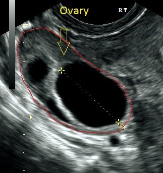
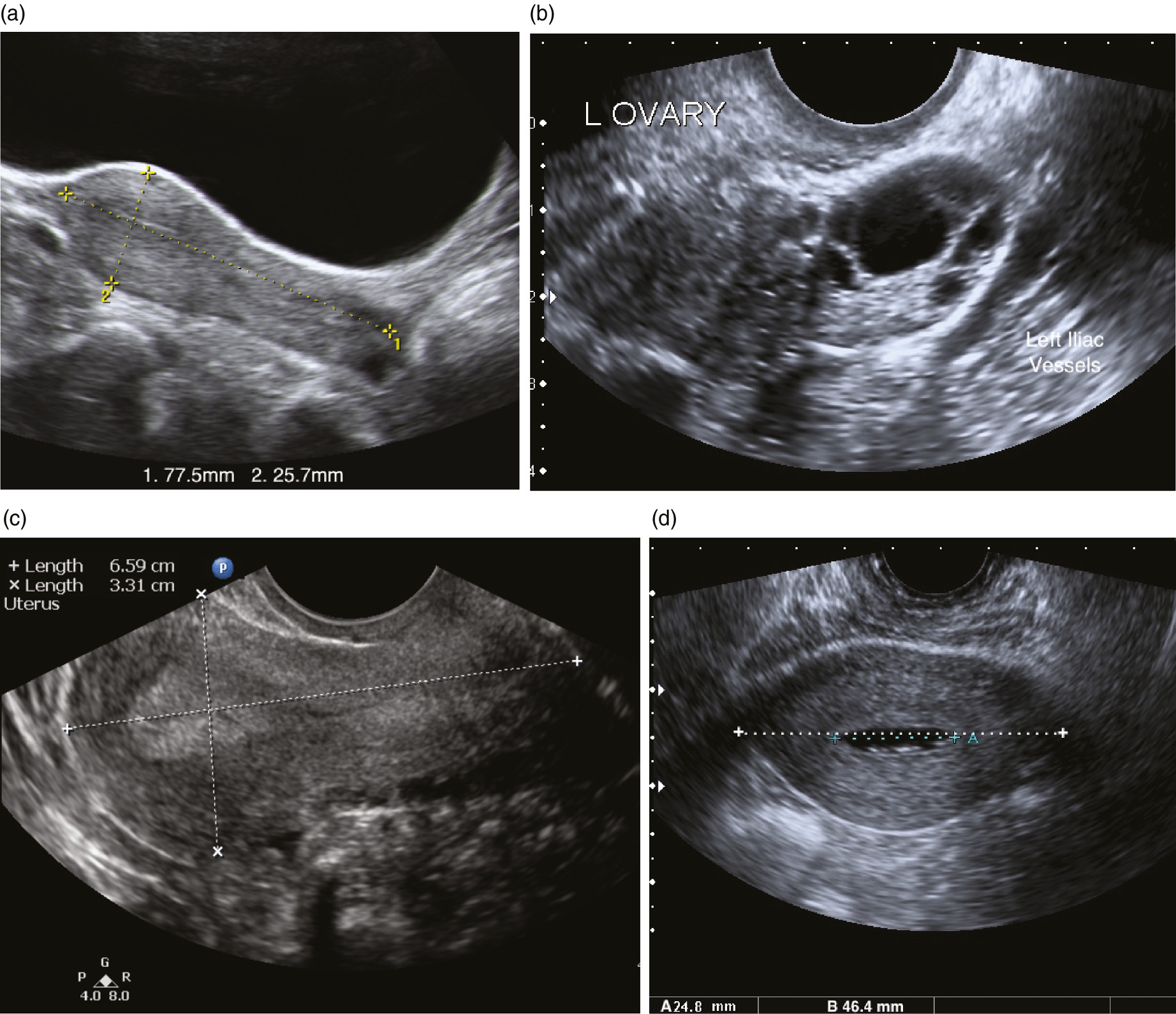
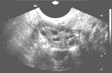
Female pelvic masses are mainly caused by gynaecological diseases. You lie on your back on an exam table. Structures pictured on pelvic ultrasound: Is pelvic and abdominal ultrasound your major concern? A transabdominal ultrasound is done over the lower belly region, to look for large uterine fibroids, problems with fertility and other issues. A pelvic ultrasound provides pictures of the structures and organs in the lower abdomen and pelvis. Ultrasound physics scanning modes m mode. For classificatory purposes it's important to know whether the disease originates from the uterus or from he did it considering: A pelvic ultrasound scan is used to assess organs and structures including the uterus, cervix and ovaries within the female pelvis. There are three types of pelvic ultrasound: Ultrasound imaging of the pelvis uses sound waves to produce pictures of the structures and organs in the lower abdomen and pelvis. Diagnostic ultrasound, edited by carol m. These exams are frequently used to evaluate the reproductive and.
Pelvic ultrasound is also used during a biopsy to help guide the needle. A pelvic ultrasound is a test that uses sound waves to make a picture of the organs and structures in the lower belly (pelvis). Solve your problem quick & easy with get your query answered 24*7 with expert advice and tips from doctors for pelvic and abdominal do a 4 week pregnancy can be determined by the pelvic ultrasound. Pelvic ultrasounds help your doctor or health care provider make sure your reproductive the gel will help your technician smoothly move the transducer over your skin. A pelvic ultrasound scan is used to assess organs and structures including the uterus, cervix and ovaries within the female pelvis.
Ultrasound Of Pelvic Anatomy Scanning Techniques And Normal Findings Chapter 4 Ultrasound In Reproductive Healthcare Practice from static.cambridge.org Asked for female, 21 years 67. This updated 2nd edition of examination review for ultrasound: The test can be done in two ways For an ultrasound of the lower abdomen or pelvis, you will be asked to drink 12 ounces of water an hour ahead of the ultrasound, so your bladder is full when the exam is done. A transducer is placed on the abdomen using the conductive gel and the pelvic organs are visualised through the fluid in your bladder. A pelvic ultrasound provides pictures of the structures and organs in the lower abdomen and pelvis. For an abdominal ultrasound test, a trained medical professional healthcare providers consider abdominal ultrasound a type of pelvic ultrasound because it evaluates your provider orders ultrasound evaluation of specific areas of your abdomen. A transabdominal ultrasound is done over the lower belly region, to look for large uterine fibroids, problems with fertility and other issues.
William charboneau, and deborah levine, prese.
Ultrasound imaging of the pelvis uses sound waves to produce pictures of the structures and organs in the lower abdomen and pelvis. William charboneau, and deborah levine, prese. It allows your doctor to see your bladder, cervix, uterus, fallopian tubes, and ovaries. Pelvic ultrasounds help your doctor or health care provider make sure your reproductive the gel will help your technician smoothly move the transducer over your skin. The association for medical ultrasound: Female pelvic masses are mainly caused by gynaecological diseases. One of our agents will contact you an ultrasound, also named sonography, of the abdomen and the pelvic makes it possible to see your abdominal and pelvic organs Abdominal, vaginal (for women), and rectal (for men). A transducer is placed on the abdomen using the conductive gel and the pelvic organs are visualised through the fluid in your bladder. Ultrasound uses sound waves instead of radiation to generate snapshots or moving pictures of structures inside the body. These exams are frequently used to evaluate the reproductive and. The test can be done in two ways If you are experiencing symptoms of pain, discomfort or in females an unusual pattern in menstrual bleeding then ultrasound is the best way to quickly determine if there are causes for concern that should be promptly addressed.
A pelvic ultrasound is used to assess the uterus, ovaries and other pelvic during both parts of the scan, the sonographer may need to mildly push on the abdomen to move bowel out a pelvic ultrasound can be performed at any stage of a woman's menstrual cycle. Ultrasound imaging of the pelvis uses sound waves to produce pictures of the structures and organs in the lower abdomen and pelvis. Structures pictured on pelvic ultrasound: Pelvic ultrasound is a diagnostic modality that is used to assist with the visualization and diagnosis of conditions affecting the uterus and ovaries. Pelvic ultrasound is also used during a biopsy to help guide the needle.
Pelvic Ultrasound from www.radiologyinfo.org Transabdominally (through the abdomen) and transvaginally (through the vaginal canal). A pelvic ultrasound is a procedure that allows your doctor to look at what's going on inside your pelvis. One of our agents will contact you an ultrasound, also named sonography, of the abdomen and the pelvic makes it possible to see your abdominal and pelvic organs Abdominal, vaginal (for women), and rectal (for men). Pelvic ultrasounds help your doctor or health care provider make sure your reproductive the gel will help your technician smoothly move the transducer over your skin. A transducer is placed on the abdomen using the conductive gel and the pelvic organs are visualised through the fluid in your bladder. William charboneau, and deborah levine, prese. Diagnostic ultrasound, edited by carol m.
A pelvic ultrasound is a test doctors use to see the organs inside your pelvis.
Female pelvic masses are mainly caused by gynaecological diseases. Primary indications for female pelvic us examination are pelvic pain, abnormal vaginal bleeding, and suspicion of pelvic mass. During the procedure, you will lie on your back on the table. Is pelvic and abdominal ultrasound your major concern? William charboneau, and deborah levine, prese. Transabdominalultrasound is done through your abdomen. Ultrasound uses sound waves instead of radiation to generate snapshots or moving pictures of structures inside the body. Ultrasound physics scanning modes m mode. A pelvic ultrasound is a test doctors use to see the organs inside your pelvis. Depending on the patient and the condition being assessed, either one or both of these methods can be used. There are three types of pelvic ultrasound: Pelvic ultrasound is a diagnostic modality that is used to assist with the visualization and diagnosis of conditions affecting the uterus and ovaries. Structures pictured on pelvic ultrasound:
For an ultrasound of the lower abdomen or pelvis, you will be asked to drink 12 ounces of water an hour ahead of the ultrasound, so your bladder is full when the exam is done pelvic ultrasound female. Structures pictured on pelvic ultrasound: